Breast Lump, is it Cancer?
Breast Lump, is it Cancer?
“Not all breast lumps cause cancer. You should know the differences between benign and malignant breast lumps.”
In this section we will discuss the following:
Breast lumps or growths found in breasts are usually not cancerous nor life-threatening. If your doctor is suspicious of a lump, he or she may need to do a biopsy to determine if it is malignant. A biopsy is a surgical procedure in which all or part of the tumor is removed. The cells from the tumor are then examined to see if they are benign or malignant.
Some types of benign lumps are:
Fibroadenomas
Fibroadenomas are benign solid tumors composed of stromal and epithelial elements. Fibroadenoma is the second most common tumor in the breast (after carcinoma) and is the most common tumor in women younger than 30 years. In contrast to cysts, fibroadenomas most often arise during the late teens and early reproductive years. Fibroadenomas are rarely seen as new masses in women after age 40 or 45 years. Clinically, fibroadenomas manifest as firm masses that are easily movable and may increase in size over several months.
Breast Cysts
Cysts within the breast parenchyma are fluid-filled, epithelial-lined cavities that vary in size from microscopic to large palpable masses containing 20 to 30 mL of fluid. A palpable cyst develops in at least 1 in every 14 women, and 50% of cysts are multiple or recurrent. The pathogenesis of cyst formation is not well understood; however, cysts appear to arise from destruction and dilation of lobules and terminal ductules
Hamartomas and Adenomas
Hamartomas and adenomas are benign proliferations of variable amounts of epithelium and stromal supporting tissue. A hamartoma is a discrete nodule that contains closely packed lobules and prominent, ectatic extralobular ducts.
Intraductal papillomas
Solitary intraductal papillomas are true polyps of epithelial-lined breast ducts. Solitary papillomas are most often located close to the areola but may be present in peripheral locations. Most papillomas are smaller than 1 cm but can grow to 4 or 5 cm. Larger papillomas may appear to arise within a cystic structure, probably representing a greatly expanded duct. Papillomas are not associated with an increased risk for breast cancer.
Sclerosing Adenosis
Adenosis refers to an increased number of small terminal ductules or acini. Adenosis is frequently associated with a proliferation of stromal tissue that produces a histologic lesion, sclerosing adenosis, which can be confused with carcinoma grossly and histologically
Fat Necrosis
Fat necrosis can mimic cancer on mammography by producing a palpable mass or density that may contain calcifications. Fat necrosis may follow an episode of trauma to the breast or be related to a prior surgical procedure or radiation therapy. Calcifications are characteristic of fat necrosis and can often be visualized on ultrasonography as well. Histologically, fat necrosis is composed of lipid-laden macrophages, scar tissue, and chronic inflammatory cells. This lesion has no malignant potential.

Malignant Breast Lumps
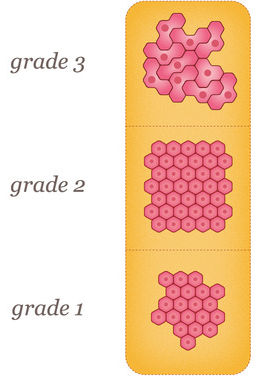
Malignant lumps or tumors are cancerous. They have the ability to invade the tissue which surrounds them. Malignant tumors are classified by grades.
If you can feel the lump in your breast, it may feel hard, or as if your breast is thicker in that spot. If you find a lump before a mammogram, it is very important that you see a healthcare professional to have it further examined. Any lump should be looked at with caution until it is determined if it is benign or malignant.
GX (No grade)
The tumor grade cannot be determined.
G1 (Low grade)
Tumor cells and tissue look well differentiated or normal.
G2 (Intermediate grade) Tumor cells and tissue look moderately well-differentiated or close to normal.
G3 (High Grade)
Tumor cells and tissue look poorly differentiated and do not look normal.
G4 (High Grade)
Tumor cells and tissue have no differentiation and do not look normal.